Prevention of Sinus Floor Augmentation
Clinical Cases
Alveolar ridge preservation in posterior maxillary teeth
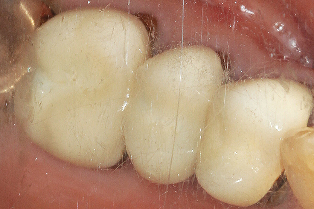
Clinical challenge
The combination of alveolar bone resorption and sinus pneumatization following tooth extraction reduces available vertical bone height for future implant placement and increases the need for sinus augmentation. Techniques for sinus augmentation such as transalveolar or lateral window approaches, which despite good predictability, are likely to cause additional patient morbidity, increase the risk of complications (e.g. sinus membrane perforation) and increase treatment costs and time.1,2 Another alternative treatment approach may involve the use of short implants (<6mm in length) in the posterior maxilla, however there is currently insufficient clinical evidence regarding its long term success.3,4
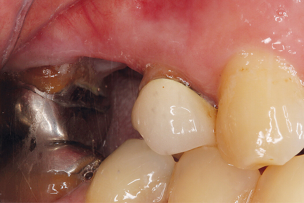
Clinical outcome at a glance
Objectives
- Alveolar ridge preservation in the posterior maxillary dentition
- Reduction of needs for sinus augmentation procedures
Conclusions
- Alveolar ridge preservation with Geistlich Bio-Oss® and Geistlich Bio-Gide® reduces the need for a sinus augmentation procedure prior to implant placement
Aim / Approach
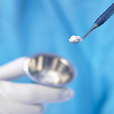
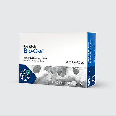
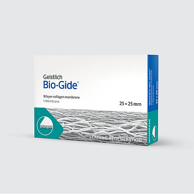
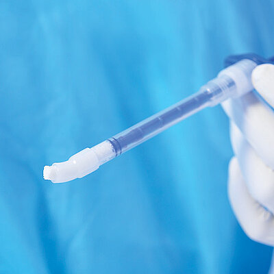
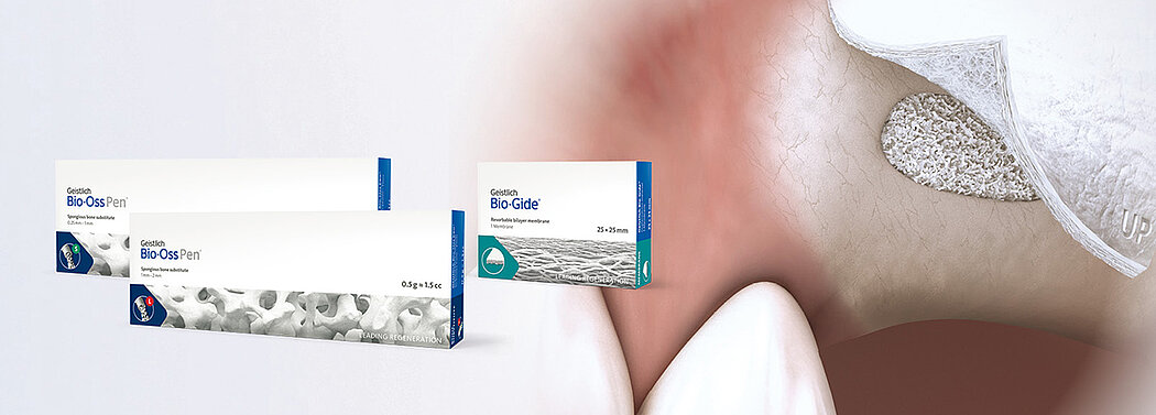
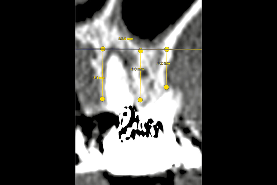
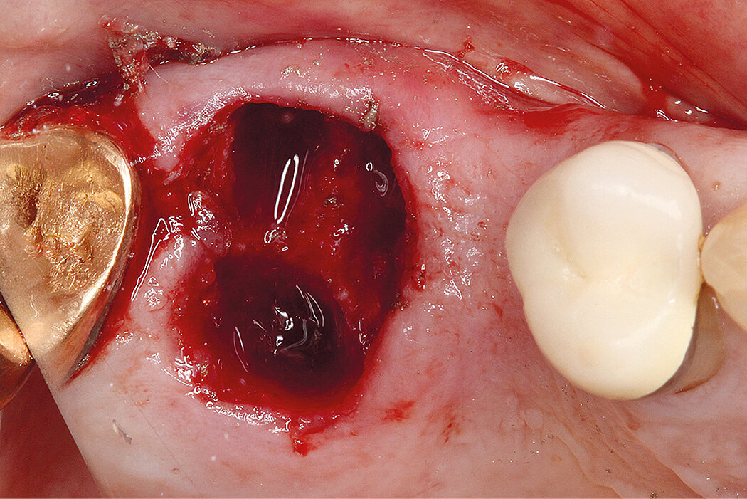
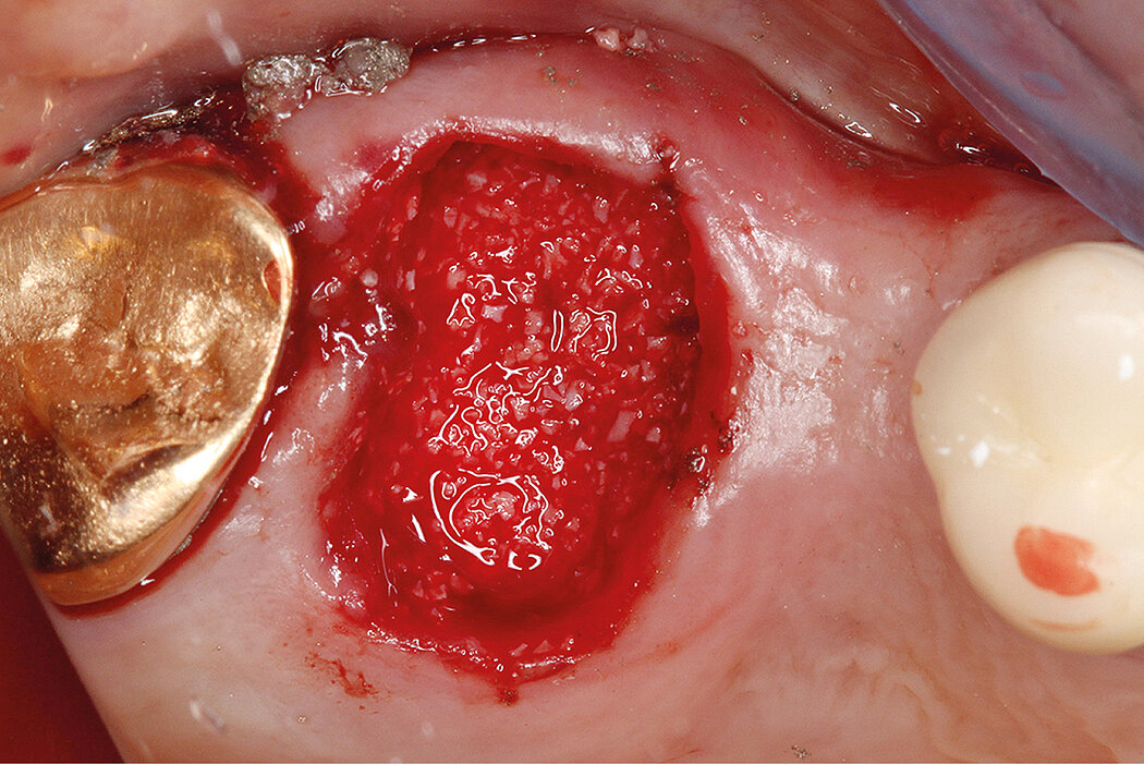
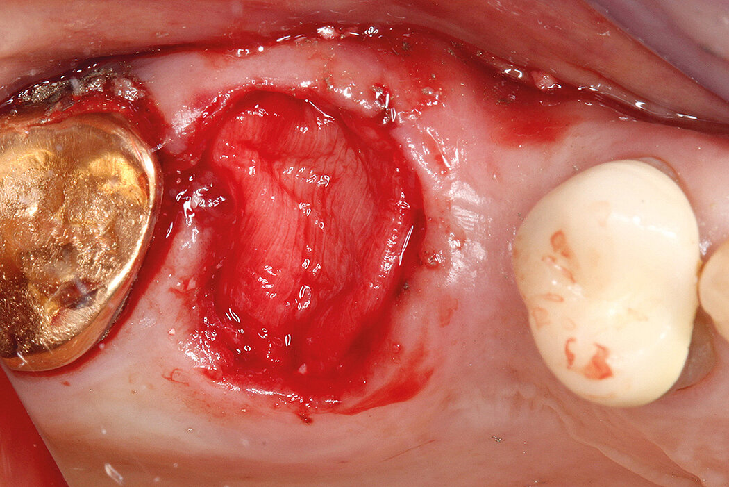
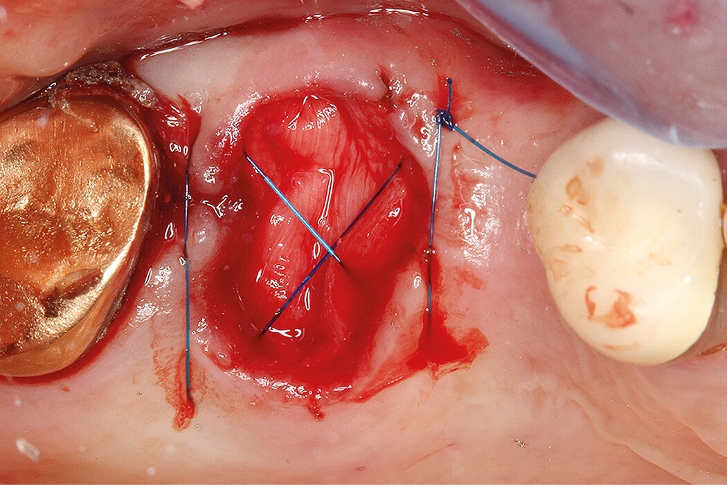
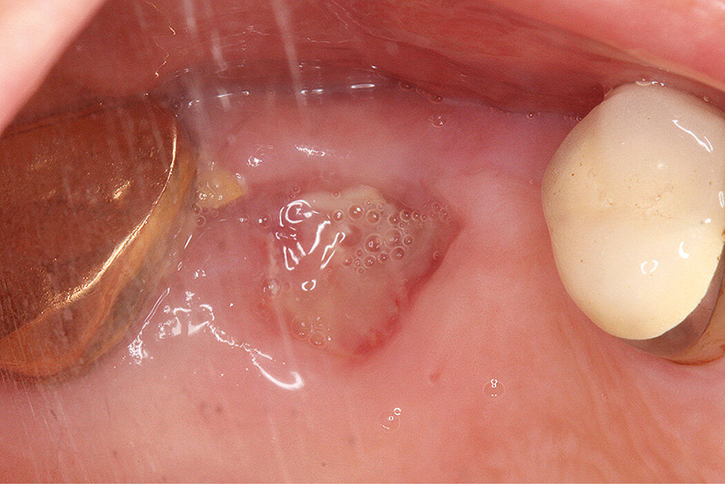
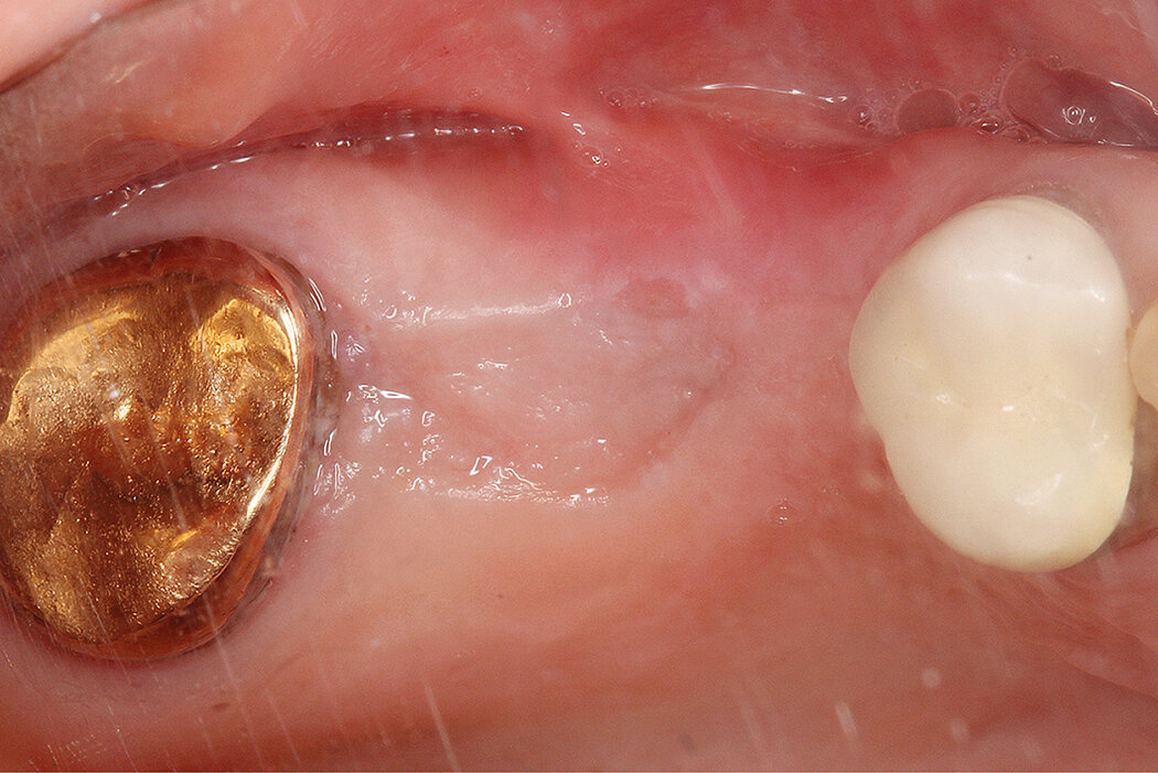
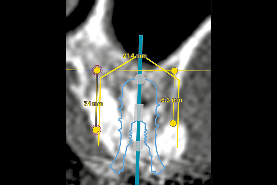
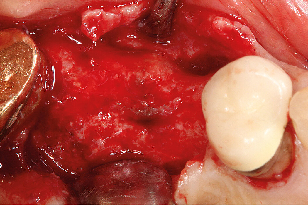
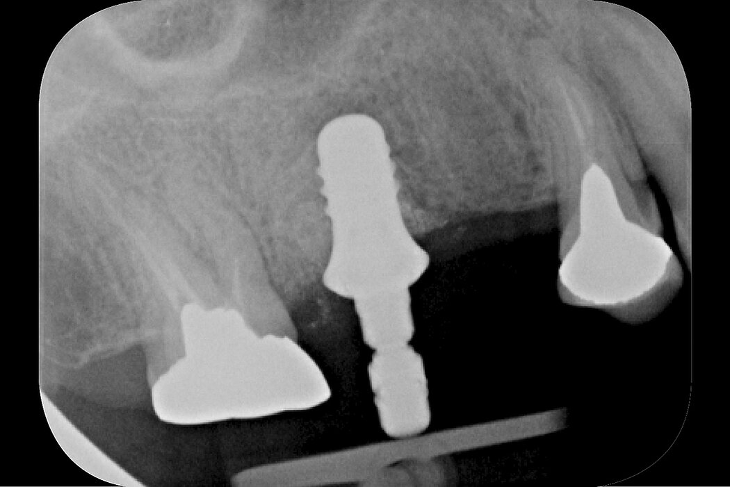
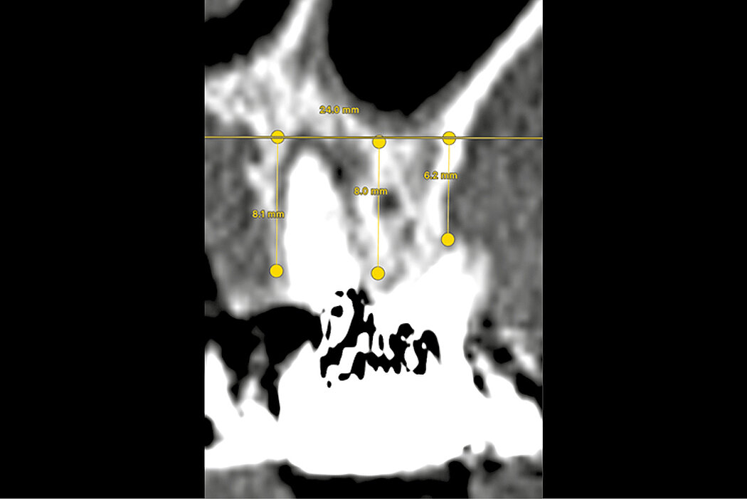
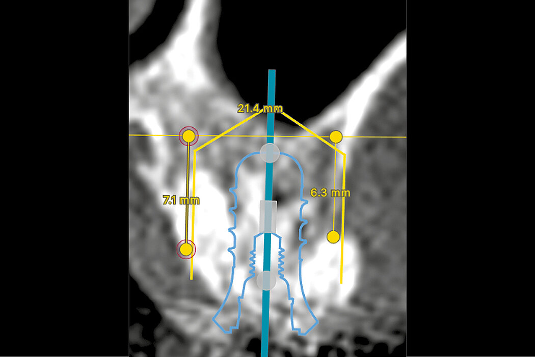
Alveolar ridge preservation in the posterior maxilla was performed to reduce the need for sinus augmentation procedures. CT scans were taken prior to extraction to assess the baseline vertical bone height. Following extraction, the socket was thoroughly debrided to remove all inflammatory or infective tissues, then, the integrity of the buccal plate was inspected. Deproteinized bovine bone mineral (Geistlich Bio-Oss®, 0.25-1.0 mm) were incrementally and firmly packed into the socket 0.5mm above the alveolar crest. Porcine collagen membrane (Geistlich Bio-Gide®) was then trimmed and covered the socket to prevent the loss of the grafted particles and provide the wound stability. Furthermore, an internal criss-cross suture5 technique was used to achieve membrane and graft stabilization without primary closure. Post-operative antibiotics and antiseptic mouth-rinses were prescribed. Sutures were removed after two weeks. After a healing period of 4 months, a clinical review and post-extraction CT scan were performed to assess any changes in vertical ridge height and sinus volume.
Conclusion
Alveolar ridge preservation following extraction of maxillary posterior teeth may minimize post-extraction remodelling and sinus pneumatization, thus reducing the need for sinus augmentation procedures prior to implant placement.
References:
- Pjetursson et al., 2008. Journal of clinical periodontology (35), 216-240 (Clinical study).
- Tan et al., 2008. J Clin Periodontol. Sep;35(8):241-54 (Clinical study).
- Fan et al., 2017. Clin Implant Dent Relat Res. Feb;19(1):207-215 (Clinical study).
- Thoma et al., 2015. J Clin Periodontol. 2015 Jan;42(1):72-80 (Clinical study).
- Park et al., 2016. J Periodontal Implant Sci. 2016 Dec;46(6):415-425 (Clinical study).